
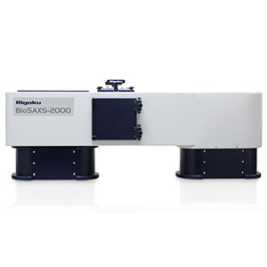
BioSAXS-2000 – Small Angle X-Ray Scattering for Structural Biology
Macromolecular crystallography and NMR are two of the most important techniques for furthering our understanding of structural biology. However, the fact remains that three-dimensional structures derived from NMR and single crystal analysis are difficult to achieve at best and quite often the analyses fail. The time, energy and money spent on cloning, expressing and purifying proteins that refuse to crystallise or are inappropriate for NMR analysis, drive up the overall cost of structural biology research and if a project is abandoned when a structure cannot be produced, the efforts are totally wasted.
SAXS Provides Complementary Structural Information to Protein Crystallography and NMR
Small Angle X-ray Scattering (SAXS) is a technique that can be applied to solutions of macromolecules to determine a variety of molecular properties, most of which are related to the shape of the molecule. The information acquired is useful both pre- and post-structure determination, making it a synergistic technique for structural biologists. You can determine the molecular weight of the molecule/complex in solution, which in turn tells you the oligomeric state in solution. You can tell if the sample is mono-disperse or aggregated. You can tell if a protein is folded, unfolded or contains disordered regions. You can determine a low resolution, three-dimensional shape of the molecule/complex, which can be used for a variety of modeling purposes.
A SAXS System Specifically Designed for Structural Biology
Rigaku’s new BioSAXS-2000 SAXS camera is designed specifically to meet the needs of the structural biologist. Based on a patented two-dimensional Kratky design, the BioSAXS-2000 takes up much less space than a conventional 3-pinhole camera but offers better flux characteristics. Best of all, the BioSAXS-2000 can be mounted on the open port of a Rigaku rotating anode x-ray generator, taking full advantage of existing infrastructure, or it can be mated to a Rigaku microfocus sealed tube X-ray source.
No Need to Wait for Beamtime at a Synchrotron
Compared to a standard Kratky camera that utilizes the line focus from a standard sealed tube source, the BioSAXS-2000 is equipped with a two-dimensional Kratky (2D Kratky) camera featuring a doubly focusing multilayer optic assembly that produces a more brilliant beam at the sample. Since the beam is focused on the detector, there is no need to de-smear the data. This design also has teh advantage over pinhole cameras in that it uses much shorter camera lengths, resulting in a higher flux. In turn, this means shorter scan times and the ability to resolve poorly diffracting structures.
Moreover, data from the BioSAXS-2000 rivals SAXS data collected at the synchrotron with data collection times usually 30 minutes or less.
Features
- Automated sample stage with 3 capillary cells and one powder standard position
- Automated sample changer (3 sample positions plus one standard)
- Simultaneous SAXS and WAXS measurements in a single image (qmax = 0.65 Å?¹)
- Easily installed on the open port of an existing generator or can be installed with your choice of X-ray source: Microfocus sealed tube, MicroMax-007 HF, FR-X
- Point focusing optics eliminates smearing issues common to traditional Kratky cameras.
- All system components are motorized for control from control computer.
- Photodiode beamstop for intensity measurements and sample absorption correction.
- Sample temperature control included with system.
- 2D Kratky collimation allows one to achieve low q measurements with no realignment
- SAXSLab data collection and processing software
System Options
- 8 or 96 position sample changer
- Automatic Analysis Pipeline (AAP) based on the world’s most popular SAXS software, ATSAS
Specifications
| X-Ray Generator | MicroMax 007HF | FR-X |
|---|---|---|
| Camera Length | ~500mm | |
| Sample Volume | 20 - 30µL | |
| Beam Size | Various width x 1500µm (at sample), 530µm (Av. at detector) | |
| Cu K alpha flux at Sample | 3.4 x 10exp9 ph/s | 8.4 x 10exp9 ph/s |
| Incident Beam Optics | OptiSAXS | |
| Collimation | 2D Kratky (X stage and tilt, motorised) | |
| Sample Stage | 3 capillaries (X,Y stage motorised) | |
| Beam Stop | PIN diode detector (motorised) | |
| q Range | 0.006 - 0.65 Å | |
| Size | 238mm (width) x 1392mm (deep) x 546mm (long), 150kg | |
| Detector DECTRIS PILATUS 100K | Sensor: reversed bias silicon diode array Active area: 83.8 x 33.5mm Pixel size: 172µm x 172µm Dynamic range: 10exp6:1 max. count rate 2 x 10exp6/sec Quantum efficiency: 99% at Cu K alpha Cooling: air coolrd |
|

 Download the AXT Life Science Product Guide