Simultaneous Correlative Light and Electron Microscopy in Diabetes Research
Diabetes mellitus is an autoimmune disease with life-threatening consequences that currently affects over 9% of the worlds population [1]. One of the two well-known forms, Type 1 diabetes, occurs when the insulin-producing beta cells are attacked and destroyed.
In the human body, these cells occur in a cluster of cells in the pancreas referred to as the islet of Langerhans. Beta cells sense the blood sugar level and release insulin when it is elevated, for example, after a meal. This in turn acts as a trigger for the uptake of glucose by the cells and its conversion to energy.
In patients with Type 1 diabetes, the beta cells are unable to perform this function, consequently retaining sugar in the blood. This could potentially lead to cell death from lack of glucose and multiple organ failure due to sustained high blood sugar levels.
At present, the cause of this condition is unknown. This study is a part of ongoing research towards gaining a fundamental understanding of this condition.
Imaging
In order to study the changes taking place during diabetes, the islets of Langerhans are imaged using electron microscopy (EM). This has the advantage of very high spatial resolution, allowing the identification of sub -cellular features. However, it is an inherently slow technique requiring scanning of the electron beam with high pixel dwell times to achieve images with a good signal-to-noise ratio. Moreover, the identification of the cells on the basis of morphology is highly labor-intensive and prone to bias.
On the other hand, fluorescence microscopy (FM) is a technique that allows the identifica-tion of cells on the basis of their function over a large field of view. Antibodies tagged with fluorophores are used as probes that attach to specific parts of the cell. The use of different fluorophores therefore allows multicolor imaging of various functionalities.
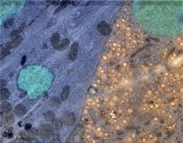
Simultaneous Correlative Light and Electron Microscopy (SCLEM) using the Delmic SECOM system combines these two methods to offer a powerful imaging technique whereby the cells of interest can be identified using FM. Subsequently, a high magnification EM image can be acquired at the desired location to reveal structural details at high resolution.
This application note describes a method for imaging the islets of Langerhans. Sample preparation details are provided and the suitability of the technique for diabetes research is demonstrated.