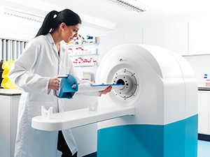
Preclinical imaging is a critical tool in the development of new treatments and also to understand the aetiology of many illnesses and diseases. It is critical that these technologies have the sensitivity and the visual acuity to match or exceed those used in a clinical setting. In a research setting the utilisation of multi-modality imaging data sets provides greater insight in the processes for the researcher.
Product Range
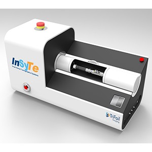
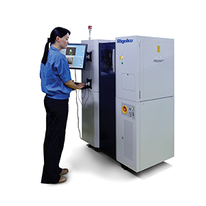
AXT now provides a range of technologies from MR Solutions, TriFoil Imaging, inviCRO and Rigaku encompassing most analytical imaging modalities. These technologies incorporate powerful analysis software as well as convenient animal handling systems to enable a fast workflow.
AXT products cater for the following imaging modalities:
Applications
Preclinical Imaging technologies are used for in vivo examination of animals to investigate changes at the organ, tissue, cellular or molecular levels. Almost all of these techniques are non-invasive.
Applications include:
- Oncology/ tumour progression studies
- Neurology
- Cardiology
- Angiography
- Embryology
- Drug development
Imaging Technologies
AXT have imaging products that employ the following technologies either singularly in combination with other technologies.
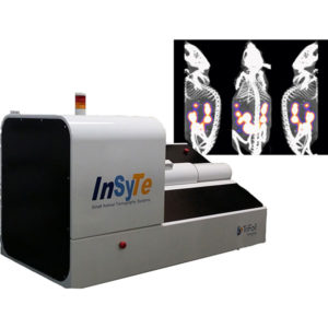
Computed Tomography (CT) |
A CT uses x-rays from a source that is rotated around the subject (or subject rotated within a stationary source) with a detector on the opposing side to the subject. The x-rays are attenuated due to variations in the density of the subject. Each rotation creates a 2D image or slice. These slices are reconstructed to form a 3D image using a sophisticated computer program.
Micro-CT has excellent spatial resolution with in vivo resolution down to 15 microns and systems like the Rigaku ano3DX which has excellent contrast can achieve resolution below 1 micron. | 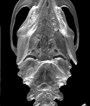 |
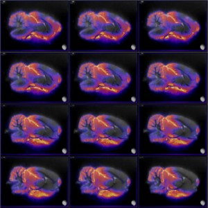
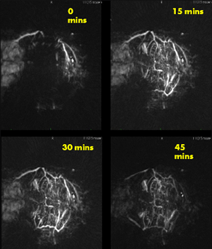
Fluorescence Emission Computed Tomography (FLECT) |
FLECT uses a laser to excite fluorophores within a specimen. In doing so, they emit light of a given wavelength that can be picked up by detectors. The detectors are rotated around the specimen and moved along its axis to provide a 360° view along the field of view.
FLECT is able to generate a full 3-D scan with uniform sensitivity and resolution throughout the entire field of view. This technique is capable of imaging deep tissue phenomena with no compromise of image quality of accuracy. | 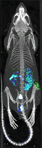 |
Magnetic Resonance Imaging (MRI) |
MRI utilises the fact that protons (water molecules) have magnetic properties and when subjected to a strong magnetic field and interrogated with an RF pulse will exhibit a signal which can be used to generate an image of the subject. An MRI imaging system consists of a powerful magnet within which the subject is placed with additional electronics and magnetic field manipulation to allow the generation of a 3D image from the subject.
MRI has excellent spatial resolution and has superior soft tissue contrast compared to CT, and as such, can distinguish between normal tissue and pathological tissue. It does not utilise ionising radiation and so is not limited by dosage restrictions. | 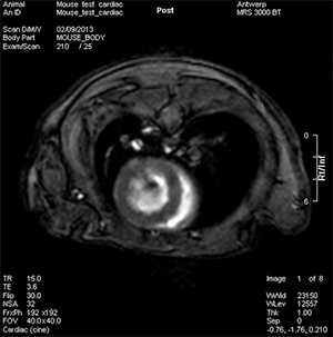 |
Positron Emission Tomography (PET) |
PET uses positron emitting radionuclides. These are usually bound to biological molecules such as 18F-FDG (F-18 Fluorodeoxyglucose) which are injected into the test subject. The PET radioactive isotopes as part of the decay process emit positrons. The positrons ultimately annihilate electrons after travelling a short distance and this process produces 2 γ-rays that are emitted 180° apart. The γ-rays are detected within a ring of sensitive detectors. The inherent geometry of this process allows the source of the emission to be localised within the subject and the subsequent data set can be used to reconstruct an image.
The high energy of the ϒ-rays means that these rays have great penetration so PET imaging has little limitation in the depth of imaging and generally acquisition time is quite short in the order of minutes. The half-life of most PET isotopes is usually quite short. F-18 for instance is 109.7 minutes. Thus ready access to a cyclotron for the production of these isotopes is a requirement. |  |
Optical Imaging |
Optical imaging techniques usually employ either fluorescence or bioluminescence:
Fluorescence is the process whereby a fluorophore is excited by light (photons) to re-emit photons of a longer wavelength. The outer shell electrons of the fluorophore move to a higher excited state due to the absorption of this light. After a short period of time these electrons drop back to lower energy state emitting a photon of a lower energy (shorter wavelength) than the exciting photon.
Bioluminescence is the process where light is generated by chemical reaction. Generally an enzyme produces light in the presence of a substrate. The best defined of these is the firefly luciferase gene.
Both of these processes (Fluorescence and Bioluminescence) are used to identify biological structures or activity. These can be employed as reporter genes (GFP or Luciferase gene) or tagged to biomolecules as a contrast agent
The detection of these agents is usually by means of a sensitive light detection system such as CCD camera. Optical systems have the advantages that they are relatively inexpensive and don’t require the infrastructure impost of nuclear imaging modalities.
The major limitation of optical imaging is the scattering of the light out of the animal tissue resulting in poor resolution usually in the mm to 10’s of mm range. The use of near infrared dyes minimises this effect. The FLECT system however, uses a sophisticated algorithm to “re-assign” the photons by modelling the scattering out of the tissue. The effect of this is to produce results similar to PET and SPECT imaging. |  |
Single Photon Emission Computed Tomography (SPECT) |
This technique is similar to PET, except it uses radioisotopes that emit γ-rays directly. The camera used to detect the γ-rays is an arrangement with a scintillation screen and sensitive light detection system. Unlike PET where the opposing ϒ-rays provide an inherent telemetry, SPECT detectors require collimation plates to determine the origin of the activity.
SPECT isotopes generally have a longer half-life than PET isotopes which lend their use longitudinal studies. Invariably SPECT isotopes such as Technicium-99 are produced at nuclear reactors although more recent cyclotrons can now produce some SPECT agents. While both have similar levels of sensitivity, SPECT has the advantage that potentially different energy radioisotopes can be conjugated to target different molecular targets allowing multiple molecular events to be imaged simultaneously. | 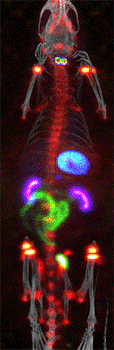 |
3D Photoacoustic CT |
Photoacoustic imaging technology combines the best features of light or optical imaging and ultrasound. This produces images that boast high optical contract and high ultrasound resolution at depth. 3D photoacoustic imaging allows volumetric images to be collected in a single snapshot. This has distinct advantages over technologies that generate 3D images slice-by-slice including:
1. The ability to capture 3D data sets at a much faster rate
2. Superior sensitivity
3. It can be used to capture dynamic (4D) photoacoustic CT scans allowing the easy quantification of probe uptake
4. It can easily determine spatial variations in probe uptake within a given volume
Furthermore, when images are collected
This unique combination makes 3D photoacoustic imaging an ideal when you are looking to generate detailed structural and functional images to give the best possible understanding of the critical disease system you are investigating.
|  |
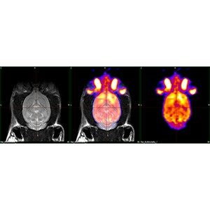
Multi-Modality Imaging |
| Molecular imaging techniques such as PET or SPECT (and FLECT) are usually combined with modalities such as CT or MRI to provide an anatomical reference. MRI is particularly useful due to its superior soft-tissue contrast. It is possible to combine multiple modalities because of the common animal bed system used by both MR Solutions and TriFoil Imaging. | 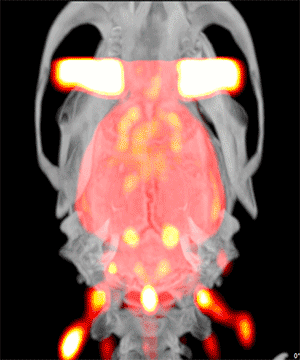 |
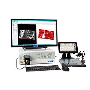
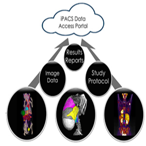
Software Solutions for Medical Research |
| inviCRO offer sophisticated software solutions to compliment AXT's range if imaging technologies. These solutions are tailor-made for translational drug discovery and development and cater for tissue to human, from target identification to Phase 3 trials across the entire electromagnetic spectrum of imaging techniques. | 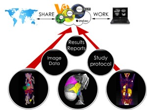 |