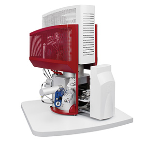
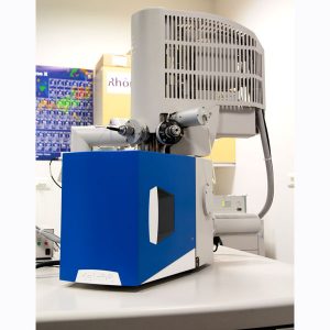
SECOM Correlative Fluorescence and Scanning Electron Microscopy
SECOM is a unique concept from DELMIC. It combines both fluorescence microscopy and scanning electron microscopy (SEM) into a single device.
Fluorescence Microscopy
For life science, fluorescence microscopy has become an indispensable research tool with its ability to map functional information. Inverted fluorescence microscopes are the favoured configuration offering superior brightness, resolution, rapid image acquisition along with flexibility of imaging and illumination sources.
The drawback with fluorescence microscopy is its inability to detect structural detail, and fluorescence data cannot be fully interpreted without corresponding structural information.
Scanning Electron Microscopy – SEM
SEM is the best tool available for imaging structural detail.
Benefits of the SECOM System
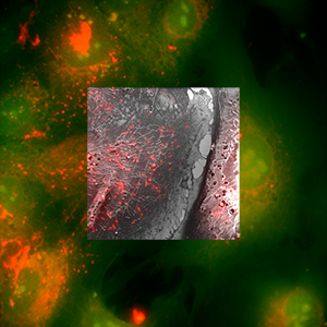
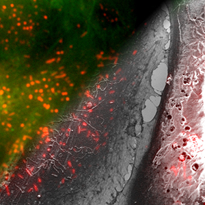
The beauty of this system is that it allows the user to obtain functional colour information via the fluorescence/optical microscope while simultaneously collecting structural information on the SEM using a single instrument. Furthermore, the ability to capture images on both system at the same time saves valuable time.
The optical axes of both microscopy systems are perfectly aligned which means that images from both techniques can be easily overlaid to yield correlative information.
Drawbacks of Conventional Techniques
While researchers have had access to both of these technologies for many years, they have never been connected as intimately as SECOM. This has meant that they have needed to be imaged in two completely separate systems. There are inherent problems working in such a manner include:
- Trying to locate the same features is an intensely laborious and time consuming task
- Many biological samples are dynamic and may undergo changes or deteriorate in the time taken between images on both system
- Moving samples from one microscope to another introduces the possibility of contamination
Features of the SECOM System
The SECOM platform incorporates a wide-field epi-fluorescence microscope. The choice of this system provides the user with the ability to obtain a direct broad overview of the sample.
Included in the package is navigation software that enables SECOM to image the same region with the SEM as was imaged with the light microscope.
Confocal Laser Scanning Microscope
The SECOM system can be optioned up with a with a Confocal Laser Scanning Microscope (CLSM). This consists of a Nikon C2+ scanhead which provides high resolution images. The high resolution images can be used to create optimal electron-optical overlays or 3D images.
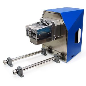
SECOM Systems
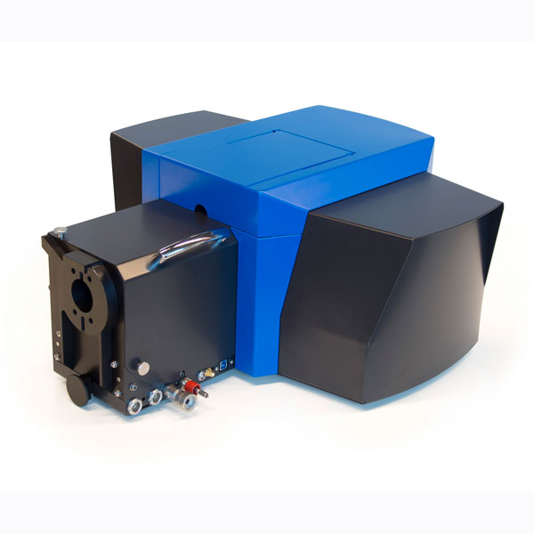
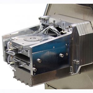
SECOM is available as a retrofit to many existing SEM systems. It simply replaces to the SEM chamber door assembly and can be installed or removed in minutes. It incorporates an inverted fluorescence microscope into the SEM chamber.
The SECOM platform includes a motorised stage and the objective light path for the optical microscope. SECOM has been designed to accommodate the same sized samples as are normally encountered in life science research.
An intuitive software interface rounds out the package and allows the user to easily acquire and manipulate images from both microscopy systems, manoeuvre the stage and control the most important settings on the SEM and fluorescence microscope.
Correlation between the two microscopy techniques is unrivalled. The alignment between the electron beam and light beam is accurate to within 0.2µm and proprietary technology allows an overlay accuracy of better than 50nm.
Aside from increased throughput with the intimate integration of the two platforms, the geometry of SECOM is fully compatible with the full range of detector modalities.
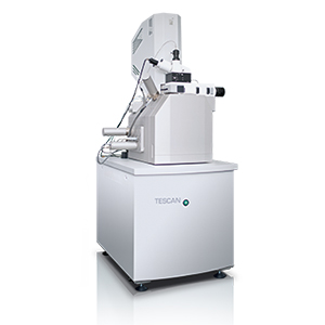
As a supplier of SEMs from TESCAN, AXT can also provide a complete turnkey system to meet your exacting requirements.
 Download the AXT Life Science Product Guide
Applications
SECOM is a versatile tool with many applications within the life science field including:
Tissue Biology and Thin Sections
SECOM is highly suited to research into tissue biology and thin sections. Fluorescence microscopy provides precise localisation of many fluorescent labels, while the SEM adds detailed structural information that exceeds results produced with traditional optical histology. Key features include:
- Suitable for large samples and array tomography
- Resolution of better then 5nm
- Multi-colour source provides compatibility with different labels
Cell Biology
Using fluorescence microscopy, researchers can gather detailed images of the intracellular distribution of fluorescent labels. SEM further enables the researcher to learn about the cell membrane configuration and structure, integrating information about morphology and distribution. Key features include:
- Detailed fluorescence information
- Easy sample navigation, guided by fluorescence
- Detailed structural information
Biomaterials Science
Imaging is a key technology used in biomaterials science and biological engineering and ties together materials science and life science. SECOM opens new doors for research in these areas streamlining the characterisation of bioengineering materials and tissues and the interactions between them as well as the effects of pharmaceuticals and foreign materials. Key features include:
- Chemical composition investigations are possible thanks to compatibility with Energy Dispersive X-Ray analysis (EDX)
- Correlation of fluorescent information and structural information relating to biometarials
- High resolution allowing visualisation of nanoparticles