SLIM Spatial Light Interference Microscopy of Live Cells and Tissues
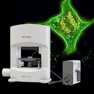
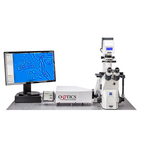
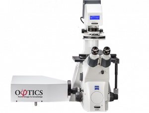
Spatial Light Interference Microscopy (SLIM) is a non-invasive, label free phase imaging technique used for imaging live cells and tissues. SLIM from Phi Optics is an add-on module for manual light microscopes that enables time-lapse imaging on a single location with single focus.
Capabilities of SLIM
SLIM upgrades the capabilities of conventional light microscopes and provides you with the following additional capabilities:
- Real-time quantitative phase imaging and display
- Non-invasive, label-free cell and tissue imaging on broad time scales (seconds to weeks)
- Programmed 2D and 3D scanning for large area 2D SLIM maps and 3D SLIM images
- Seamless overlay with other microscopy channels
- Quick and easy segmentation of cells
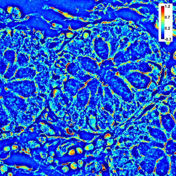 | 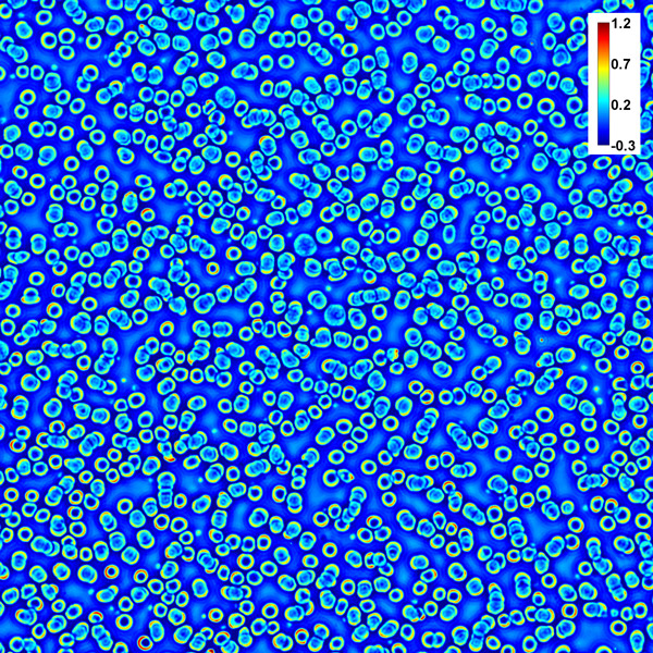 | 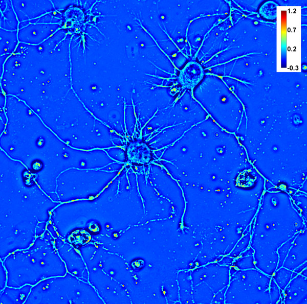 |
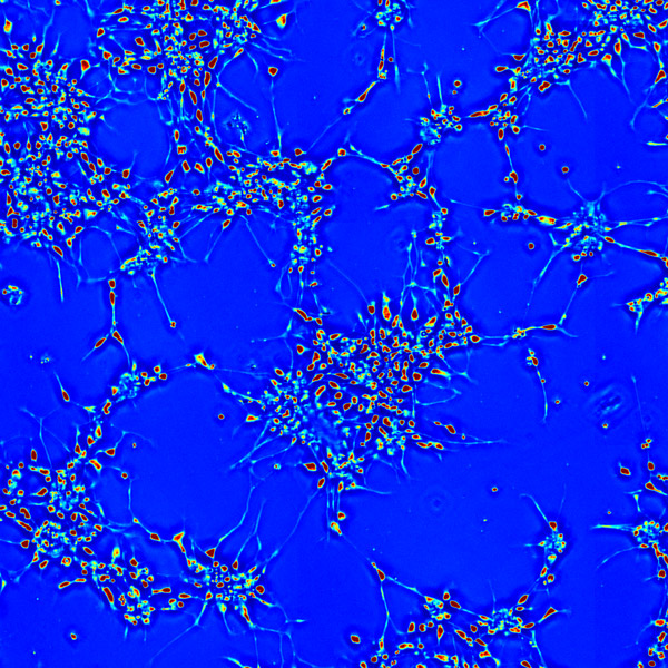
| SLIM image of a prostate tissue biopsy (100x 1.3NA objective). | SLIM image of blood smear (40x 0.65NA objective). | SLIM image of rat hippocampal neurons (40x 0.65NA objective). |
What is SLIM?
SLIM is a wide field imaging technology that is suitable for simultaneously measuring large cell populations at full camera resolution e.g. 2mm FOV for 10x objective with 4.2MP camera. 3D tomography is also possible using wide field optical sectioning (e.g. 850nm Z-resolution for 100x/1.4NA objective).
The same camera is used to acquire all microscope outputs. This means that SLIM and fluorescence images can be seamlessly overlayed.
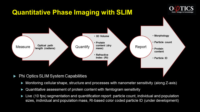
How Does SLIM Work?
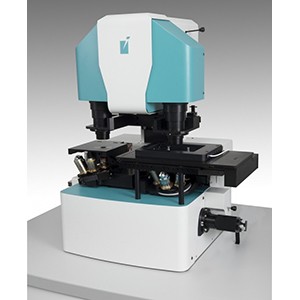
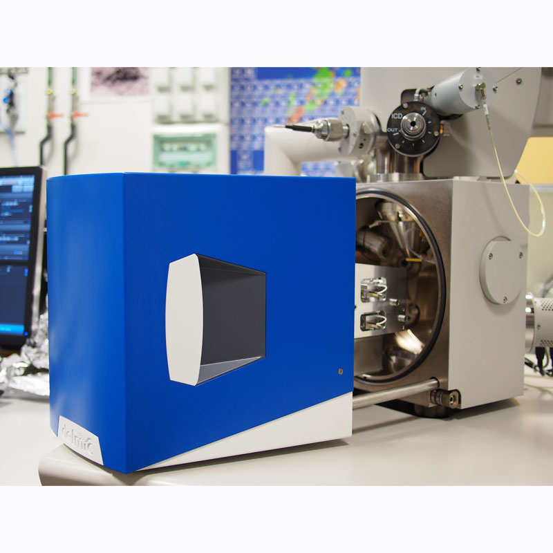
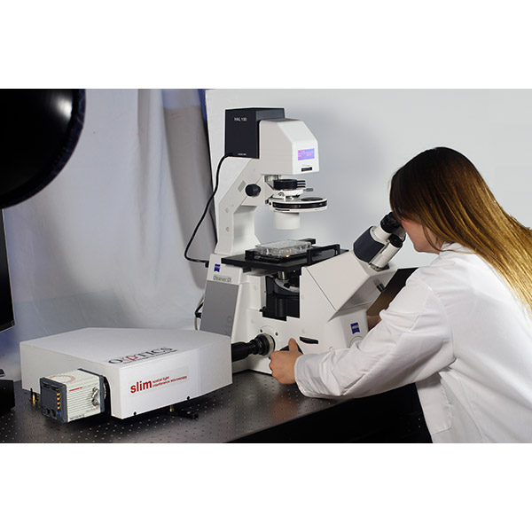
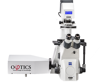
SLIM uses optical interferometry which is extremely sensitive to structure and dynamics. The Phi Optics SLIM systems is an add-on compatible with all major microscope brands (10x to 100x magnification) and overlays with fluorescence. It connects via a C-mount and uses the microscopes white light illumination source.
Optical interferometry phase imaging quantifies the optical path length difference in a biospecimen and converts this to thickness, dry mass area density and refractive index maps.
When a live cell in culture medium is imaged using microscopes phase contrast mode, the light that passes through the object (scattered beam) and the light that passes though the medium (reference beam) combine through interference in the image plane. The shift in optical paths (or phase shift) between the two beams is measured and is proportional to the optical density at that point, with denser areas e.g. nuclei resulting in a larger phase shift.
The SLIM module relays signals from the image plane at a 1:1 ratio to a CCD camera at its exit port. At the heart of the SLIM module is a liquid crystal spatial light modulator (SLM). It is conjugated with the back focal plane of the microscope objective and modulates the reference beam akin to a phase plate with variable thickness. Using a tunable phase ring and shifting the phase of the reference beam by fixed amounts (0, 0.5?, ? and 1.5?) the camera captures frames and creates a quantitative phase image by combining the four images and solving the field interference equation.
 Applications
SLIM is suited to a range of applications in the life science arena including:
- Cell growth
- Cell dynamics
- 3D tomography
- Neuroscience
- Blood testing
- Tissue imaging
- Drug discovery
- Embryo viability
- Stem cell manipulation
- Digital pathology
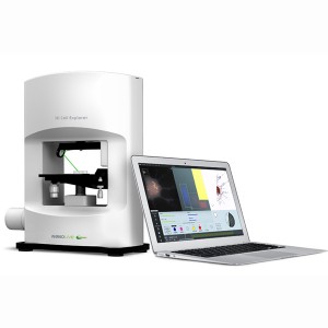
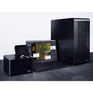
Models and Specifications
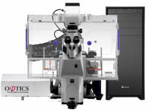
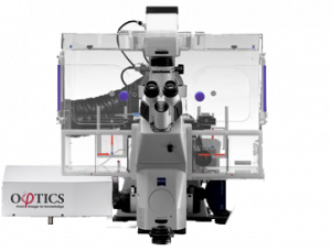
| SLIM ULTIMATE | SLIM PRO | SLIM BASIC | |
|---|---|---|---|
 |  |  |
|
| Real Time Imaging | Yes | Yes | Yes |
| Programmed 2D Scanning | Yes | Yes | No |
| Programmed 3D Scanning | Yes | Yes | No |
| Programmed Fluorescence Overlay | Yes | Yes | No |
| SLIM Acquisition Speed | Up to 12 fps (2048 x 2048) | Up to 4 fps (1928 x 1448) | |
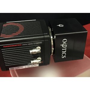
| Camera | Hamamatsu ORCA-Flash 4.0V3, 2048 x 2048 Resolution, 100 fps, 16 bit | Point Grey USB CCD, 1928 x 1448 Resolution, 26 fps,14 bit | |
| Computer & Storage | Intel Core i7, 32GB RAM, 2TB SSD + 100TB HDD, NVIDIA GeForce GTX 780, Windows 7 | Intel Core i7, 16GB RAM, 1TB HDD, NVIDIA GeForce GTX 780, Windows 7 | |
| Software | Acquisition: CellVista Pro Postprocessing: ImageJ Phi Optics Toolbox | Acquisition: CellVista Basic Postprocessing: ImageJ Phi Optics Toolbox | |
| Hardware Footprint (L x W x H) | 43 x 36 x 11 cm | ||
| Measurement Resolution | Path Length Sensitivity <1nm Transverse Resolution – diffraction limited (e.g. 350 nm with 63x/1.4NA objective) Axial Resolution – optical sectioning (e.g. 890 nm for 63x/1.4NA objective) |
||