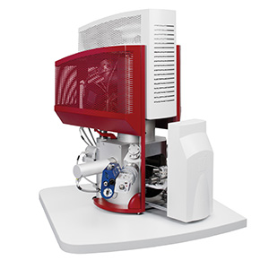
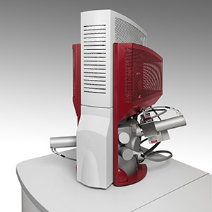
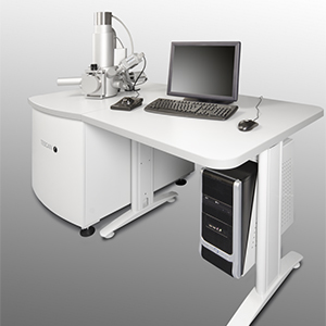
VEGA SB – Full Featured SEM for the Price of a Bench Top SEM
The TESCAN VEGA SB is a full-functioning SEM for the price of a bench top SEM. It offers all the functionality of a limited bench top system plus more! It makes the perfect system for teaching with its excellent ease of use and extremely low maintenance requirements. Moreover, it is also a proven performer in the field of research where many prestigious institutions have used it to generate publications quality data.
Perhaps the biggest advantage of this system compared to benchtop SEMs is that, it can grow with your increasing demands of your laboratory. It offers the flexibility to integrate a range of detectors and accessories (up to 10) to further increase its analytical capabilities.
The current VEGA SB is the third generation, benefitting from 12 years of continuous development. Areas that have benefitted from the latest technological developments providing performance exceeding those of bench top systems and equal to more expensive systems include:
- Improved high-performance electronics to offer the fastest scan rate and image acquisition
- TESCAN unique 4 lens column offering 5 imaging modes including wide field and channelling
- In-Flight Beam Tracing for accurate and live probe current/spot size adjustment
Despite the technology upgrades, the VEGA maintains an excellent price to-performance ratio, making it an excellent value proposition for your lab.
As with other TESCAN VEGA systems, it utilises a conventional tungsten emitter and can operate in either high or variable pressure modes, making it suitable for all materials and life science applications.
Analytical Features
To give your system analytical power and flexibility the VEGA SB includes:
- 5-axis eucentric sample stage (X, Y, Z, tilt and rotation)
- First-class YAG scintillator-based detectors
- A range of optional detectors and accessories
- Rapid chamber evacuation using turbomolecular and rotary vacuum pumps
- Ability to image non-conductive samples using variable pressure mode (UniVac)
- Ability to have STEM and EBIC detectors
- A dedicated SE detector for high pressure operation (LVSTD)
- Ability to perform CL, EDS, EBSD, EBL and more
Ease of Use and Automated Functions
To make SEM analysis as easy and pleasurable as possible, so you can spend more time analysing and less time setting up, the TESCAN VEGA system uses a fast and intuitive software interface to control all functionalities. This interface is suited to users of all experience levels and will enable even novice users to be imaging samples in the shortest timeframes. It includes such things as self-diagnostics for system readiness checks, image management and report.
To further enhance the user experience, many functions are automated such as:
- Filament heating
- Gun and column alignment
- Focusing, astigmatism correction and brightness/contrast adjustment
The remote control interface also allows many of the microscopes features to be controlled from another PC not directly attached to the system.
Optics
The VEGA SB benefits from the following:
- Continuous adjustment of the beam energy from 200 V up to 30 kV
- High beam current for analytical and EBL works (up to 200 nA)
- TESCANs unique four-lens Wide Field Optics incorporating their proprietary Intermediate Lens (IML) providing a range of working and display modes as well as enganced field-of-view and depth of focus
- State-of-the-art scanning coils and electronics enable an ultra-fast imaging rate, down to 20ns/pixel with minimal dynamic distortion effects
- Real Time In-Flight Beam Tracing for high-precision real-time computation of optics parameters
- Fully automated column design with no mechanical centring elements
- Unique live stereoscopic imaging using advanced 3D beam Technology
Extremely Low and Easy Maintenance
The VEGA has been designed with ease of maintenance in mind so it is easier for you to keep your instrument in peak operating order. This allows you to spend more time collecting data and minimises downtime (less than 5%).
Optics
| Property | Value | |
| Electron source | Tungsten heated cathode | |
| Magnification | 3X to 1,000,000X (for 5” image width in Continual Wide Field/Resolution) | |
| Accelerating voltage | continuously adjustable between 200V to 30kV | |
| Resolution SE detector | 3 nm @ 30 kV | 8 nm @ 3kV |
| Resolution BSE and LVSTD detectors | 3.5 nm @ 30 kV | |
| Max. field of view | 24mm at WD 30mm | |
| Probe current | continuously adjustable between 1pA to 2µA | |
| Electron optics working modes | - Resolution – High-Resolution mode - Depth – Sets the column up in a mode that enhances depth of focus - Field – Optimises the column to provide a large non-distorted field of view - Wide Field – Provides an extra large non-distorted field of view for extra low magnification imaging - Channeling - Working mode for assessment of crystal orientation data of the specimen, acquiring of electron channeling pattern (ECP) |
|
Scanning
- Scanning Speed – From 20 ns to 10 ms per pixel adjustable in steps or continuously
- Scanning Features – Point & Line Scan
- Focus Window shape, size and position continuously adjustable
- Dynamic Focus in plane or folded plane tilted up to ±70 deg
- Image rotation, Image shift, Tilt compensation
- 3D Beam defined tilting scanning axis around XY axis
- Live Stereoscopic Imaging
- Other scanning shapes available through optional DrawBeam software
Chamber
| Internal size | 160mm dia |
| Door | 120mm (width) |
| Number of ports | 10 |
| Chamber suspension | Mechanaical |
Specimen Stage
| Type | Eucentric |
| Movement | X=45mm (motorised) Y=45mm motorised) Z=27mm, Z’=6mm Rotation=360° (motorised), Tilt=-90° to +90° |
| Specimen height | 36mm max. |