
X-Ray Generators and X-Ray Sources
Rigaku is a world-leader in analytical x-ray technology and the design and manufacture of advanced x-ray generators is one of their core competencies. These can be purchased as stand-alone items for custom applications or where Rigaku does not have a bespoke system.
![]()
![]()
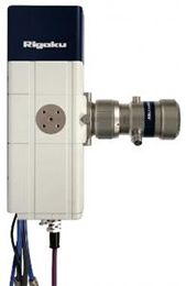
The Rigaku range includes high power rotating anode x-ray generators, sealed tube sources as well as compact x-ray tubes from Newton Scientific.
FR-X Ultra-High Intensity Microfocus Rotating Anode X-Ray Generator
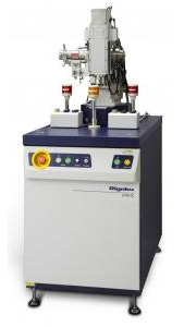 Designed for structural biology and materials science, the FR-X boasts the highest usable x-ray flux for laboratory applications. The x-ray flux is up 20% on the previous model with a new design featuring direct-drive anode for reduced maintenance and running costs. The dual port design with dual wavelength anode provides the ultimate in flexibility.
The design of the FR-X x-ray generator benefits from decades of proven craftsmanship so you can be guaranteed reliable, dependable, high powered (3kW) performance.
Features
- Highest flux of any microfocus rotating anode source
- Direct-drive for reduced maintenance and running costs
- Smallest focal spot size of any rotating anode generator at 70µm
- Small beam provides greater flux density and lower background
- High flux equates to reduced exposure times and higher throughput
- Collect full data sets on samples where only low-resolution reflections were seen on a standard system
- Screen crystals where no diffraction is seen using a lower power systems
- Optional dual wavelength anodes with computer controlled wavelength selection
- Choice of anode materials: Cu, Cr, Mo, Cu/Cr, Cu/Co
MicroMax-007 HF Microfocus Rotating Anode X-Ray Generator
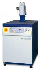 The MicroMax-007 HF is the most commonly used homelab x-ray source for protein crystallography applications and is also popular for small molecule crystallography. With a sub-100µm focal spot size, it offers high brilliance that can be used effectively to illuminate small crystals. The latest HF variant offers a 50% increase in power loading that rivals second generation synchrotron sources, with the advantage of increased beam stability and access.
This system was designed with optimal up-time in mind. The design also enables quick and easy filament changes using a unique filament cartridge that take about 45 minutes.
Other design features include a direct-drive anode and compact tower assembly that houses the vacuum chamber and turbo-molecular pump for rapid pump downs. The tower is mounted on the generator tabletop and can be easily moved for integration with other x-ray optics.
Features
- Smallest focal spot size of any rotating anode generator at 70µm
- 1.2kW of power in a small beam providing high flux density and less background
- Unique pre-aligned and pre-crystallised filament cartridges minimising maintenance
- Easily manoeuvrable anode for optimal experimental configuration
- Anode materials Cu, Cr, Mo and Co
- 2 ports for increased versatility
- Extended anode and filament lifetimes
MultiMax-9 9kW Multi-Purpose Rotating Anode X-Ray Generator
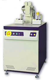 The MultiMax 9 is a versatile, compact and powerful (9kW), stand-alone x-ray source that can be adapted to many different applications. It is based on proven technology, so reliability is ensured.
Features
- High brilliance 3.6kW/mm2
- Compact design with small footprint
- Adjustable tube tower height
- Line and point focus modes
- Low cost of ownership
- Anode materials Cu, Cr, Fe, Co, Ni, Mo, Au, Ag and W
Line Focus Applications
- Powder diffraction
- Pair distribution function
- Direct replacement of redundant systems
Point Focus Applications
- Laue diffraction work
- Guinier camera work
- Diamond anvil cells
- Small molecule crystallography
- Direct replacement of redundant systems
ultraX 18 18kW Rotating Anode X-Ray Generator
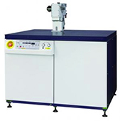 The ultraX 18 is a powerful, high-frequency x-ray generator rated to 18kW. It has been designed for flexibility, high-stability and low maintenance operation. Variable anode mounting orientations allow the system to produce two point focus beams or two line focus beams, while the anode housing itself can be mounted in any position.
Features
- Powerful x-ray generator producing either point of line focus beams
- The x-ray tube, high-voltage connection unit, and main vacuum system are compactly assembled on the top of the worktable
- Easily adjustable x-ray tube position permits different optical configurations
- The compact tube housing allows the close locating of the optical system and the x-ray source
- The X-ray tube in one compact body permits rapid start-up of the entire system
- Anode materials: Cu, Cr, Fe, Co, Ni, Mo, Ag, W and Au
- An ideal high flux generator option for small molecule systems and many XRD applications
MicroMax-003 Microfocus High-Intensity Sealed Tube X-Ray Source
 The MicroMax-003 offers of a microfocus x-ray source with low power consumption and long life, with minimal maintenance.
The system incorporates a tightly coupled microfocus generator and specially designed confocal multilayer optic that result in a system requiring an absolute minimum of maintenance.
The MicroMax-003 can be easily integrated into numerous different x-ray systems and offers excellent reliability and uptime.
Features
- Very small beam, <100µm at the crystal provides superior signal to noise
- Integrated optics optimised for HPAD (Hybrid Pixel Array Detectors) and IP (imaging plate) detectors
- Confocal MaxFlux double bounce optics provides up to 70 times better Cu Kß suppression than single bounce optics. This produces a beam that has less noise, lower divergence and increased intensity, making it ideal for small crystal samples
- 3 year tube warranty
Sealed Tube X-Ray Sources from Newton Scientific
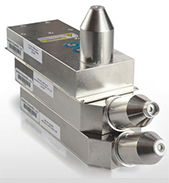 Newton Scientific is a high voltage technology company that manufactures miniature x-ray sources. Their precision monoblock sources are ideal for applications that require small low power consumption x-ray sources such as handheld, portable or benchtop systems. They feature miniature sealed tubes with transmission type end windows with built-in high voltage power supply, all housed in a radiation-shielded unit.
The range includes a number of standard designs, while custom designs are also available to suit your particular application.
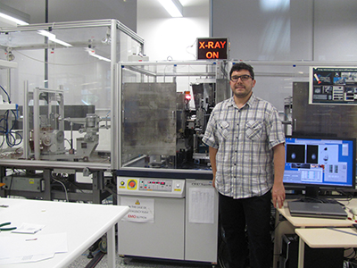
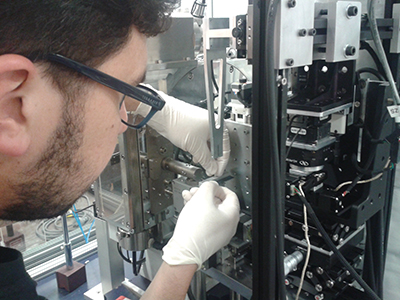
Australias Most Versatile X-Ray Instrument Based on Rigaku Rotating Anode Source
When people think of x-rays as a source for analytical applications, most of us think of instruments dedicated to a single technology like XRF or XRD and variations thereof. However, housed in the School of Physics and Astronomy at Monash University is a lab run by Dr. Daniele Pelliccia. Over the last 24 months, he has been building a unique facility with a raft of different capabilities, really exploiting the analytical power of x-rays and pushing the bounds of existing technologies.
 Bringing his years of experience working with synchrotrons, Daniele came to Monash University in 2009. In 2012 he was awarded a Discovery Early Career Research Award by the Australian Research Council and received the Monash University Research Accelerator Award which has allowed him to build this facility.
At the heart of his system is a Rigaku FRE+ Superbright x-ray generator, featuring rotating anode technology. When asked why he chose the FRE+ Superbright Daniele said, The choice was simple. At the time of purchase it was the brightest microfocus x-ray source on the market with the smallest focal spot of any rotating anode generator source providing excellent flux density, making an excellent starting point for our project. While some people questioned the reliability of the rotating anode, I can say this is a myth if we adhere to the recommended maintenance schedules
Furthermore, the FRE+ x-ray generator has two ports that effectively provide two beam lines, said Daniele. To these, he has designed and built several custom instruments that allow him to carry out a range of different analytical techniques, with both beam lines being available for simultaneous use.
Beam Line 1
- Diffractometry (XRD) – Including standard techniques such as powder diffraction, single crystal and stress analysis
- X-ray reflectivity (XRR) For thin films interfaces or multilayers characterisation
- Ultra-small angle x-ray scattering (USAXS) For characterisation of micro and nanostructures on surfaces, or dispersed in solution
Beam Line 2
- X-ray microscopy (XRM) 1µm resolution x-ray imaging of small samples (2-3 mm thick)
- X-ray micro-CT Similar to XRM with 3D reconstruction
- Small and wide-angle scattering (SAXS and WAXS) – For nano-structural characterisation (up to about 20° collection angle)
This variety of techniques allows his system to cater for both materials and biomedical characterisation, two vastly different fields of research.
Using x-ray microscopy, Daniele is looking at biological systems, imaging living cells and small biological systems. More specifically, he is looking at imaging muscles in Zebrafish which are commonly used in the research of human diseases. Currently, muscles can only be viewed using optical fluorescence microscopes with the aid of stains which can cause distortions. X-ray microscopy will avoid the need for staining providing natural contrast for different tissues.
The goal of this area of research is two-fold: producing high-quality images of biological systems for biomedical research and the development of a benchtop sized x-ray microscope.
In the realms of materials science, Daniele is carrying out nanodiffraction experiments to measure the properties of nanomaterials. By using x-ray scattering, the size of the particles can be measured indirectly, providing information about their size, shape and orientation. Further developing this system will enable applications in nanotechnology, nanosafety and materials science. Concurrently, Daniele’s team is collaborating with Monash materials researchers to develop lighter, stronger metals reinforced with nanoparticles.
 These two areas of research could collide with Daniele also interested in looking at the potential toxicity of nanoparticles on the human body, an area that has not kept pace with the commercialisation of nanomaterials, and hence in need of improved scientific understanding.
Daniele and his team have already carried out cutting edge research in x-ray optical physics and phase contrast imaging, resulting in a number of publications, some of which have been published in the refereed/open access journals Optics Express and Biomedical Optics Express. Titles include:
- Multi-modal hard x-ray imaging with a laboratory source using selective reflection from a mirror
- Dual scanning and full-field hard x-ray microscopy with a laboratory source
- X-ray phase imaging with a laboratory source using selective reflection from a mirror
They are also collaborating with researchers form other institutions such as Deakin University. In this example, they are using SAXS and WAXS to delve into the nanostructure of carbon fibres to try and optimise processing and properties.
It is great to see the level of innovation, expertise and dedication that Daniele and his team have in this project as well as the support from the Australian Research Council and Monash University to enable them to create such a unique facility in Australia.
Publications
Some key publications derived from Daniele’s work:
- D. Pelliccia and D.M. Paganin, X-ray phase imaging with a laboratory source using selective reflection from a mirror, Opt. Express, Vol. 21, Issue 8, pp. 9308-9314 (2013).
This paper was selected by the editor and featured in a special edition of “Spotlight on Optics” which highlight important papers published in the journals of the Optical Society of America. The accompanying review to the paper in plain english is available here: http://www.opticsinfobase.org/spotlight/summary.cfm?uri=oe-21-8-9308&ori
- C. J. Blackhall, K. S. Morgan and D. Pelliccia, Dual scanning and full-field hard x-ray microscopy with a laboratory source, Opt. Express Vol. 22, pp. 15437-15446 (2014).
- D. Pelliccia and D.M. Paganin, Multi-modal hard x-ray imaging with a laboratory source using selective reflection from a mirror, Biomed. Opt. Express Vol. 5, pp. 1153-1159 (2014).
Australian Researcher Builds Multi-Modal X-Ray Microscope and Micro-CT with Bragg Magnifier
Dr. Daniele Pelliccia from RMIT University has built Australias most versatile X-ray instrument. In conjunction with Patrik Vagovic and other researchers from Germany and the Slovak Academy of Sciences , Daniele has recently published a paper entitled Laboratory-Based Multi-Modal X-ray Microscopy and Micro-CT with Bragg Magnifier in the journal Optics Express (DOI: 10.1364/OE.23.018391). Using novel Bragg magnifier optics, the researchers have been able to combine different microscopy modalities together into a single instrument using a lab-sized X-ray source
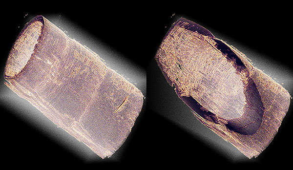
Figure 1. Selected 3D renderings of the micro-CT of the antenna of a tiger prawn. The specimen is about 1 mm in diameter and produces a significant attenuation for X-rays. Micro-CT enables non-destructive inspection of sample volumes. In this example both outer and inner walls as well as he structure of the antenna wall itself can be studied through virtual cuts across the rendered volume.
as opposed to resorting to synchrotron radiation.
At the heart of the system is a Rigaku FRE+ rotating anode X-ray generator high brightness X-ray source. Out of the box, the FRE+ has a focal spot size in the order of 80 to 100µm and is not generally suited used imaging applications. However, Daniele and his colleagues designed a novel optics system that magnifies the X-ray beam acting as X-ray zoom lens, resulting in an increase in resolution. While the optic is somewhat complex, it actually adds an additional level of flexibility to the system.
The optic itself consists of two channel-cut crystals in asymmetric diffraction. This produces an X-ray beam that is magnified by 15X and highly collimated that is suited to high-resolution imaging. Furthermore, thanks to the Rigaku FRE+, the system is suited to imaging weak, radiation damage sensitive samples. The Bragg magnifier optic is able to generate quantitative data for analyses such as:
- Microstructure determination
- 3D Computed Tomography or micro-CT
- Small angle scattering directionality
It performs these functions being sensitive to X-ray attenuation, refraction as towel as X-ray scattering. Due to the fact that the Bragg Magnifier requires an intense X-ray beam to perform, a sealed tube type X-ray source is simply not powerful enough, nor is it able to generate high enough sensitivity.
The system is also able to operate as a multi-modal X-ray microscope. In this guise, it can image in both bright field and dark field modes, which would not be possible using a sealed tube X-ray source. This imaging mode is somewhat similar to an optical microscope and is capable of revealing high levels of detail thanks to the large variations in contrast that it can produce.
In micro-CT mode, the system is able to image samples with a 5µm resolution, which rivals other commercially available systems.
In summary, the novel optic developed by these researchers has been able to extend the imaging capabilities of the high intensity X-ray source like the Rigaku FRE+ with large focal spot to create a high resolution X-ray microscope and micro-CT. The high flux rotating anode X-ray source is key to the system, with sealed tube X-ray sources not able to produce sufficient X-ray flux to efficiently use innovative optics to image specimens. This new experimental setup effectively brings synchrotron micro-imaging capabilities within the reach of laboratory researchers.
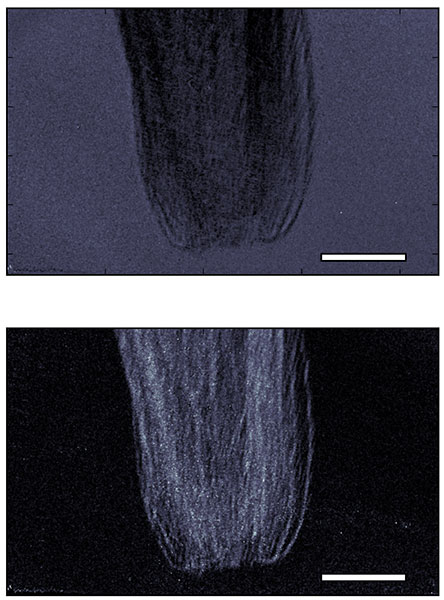
Figure 2. Top: bright field image of the tip of a dandelion seed. The image corresponds to the attenuation of X-rays in the sample. Bottom: Dark-field image of the same specimen. The dark field contrast arises from X-rays that are being scattered in the horizontal direction (Small-angle X-ray scattering). The scale bar corresponds to 150 microns.
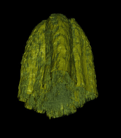
Figure 3. Volume rendering of the micro-CT of the dandelion sample.