
Lyncean Compact Light Source at TUM Facilitates Groundbreaking Biomedical Imaging Research
 The Munich Compact Light Source (MuCLS), developed and manufactured by Lyncean Technologies and subsequently installed at the Munich School of BioEngineering, on the campus of The Technical University of Munich (TUM), is being utilised for groundbreaking biomedical imaging research. Three recent publications highlight how the quasi-monochromatic, energy tunable, partially coherent light generated by the MuCLS can be used to increase contrast in techniques that can be applied to real world medical diagnosis problems.
The Munich Compact Light Source (MuCLS) is a mini synchrotron capable of creating a nearly monochromatic, energy tunable X-ray beam. X-rays are generated via inverse Compton scattering when an electron bunch circulating at 65 MHz in a miniature storage ring collides with high power laser pulses stored in a resonant optical cavity. This new generation X-ray source is able to deliver synchrotron-quality data for a wide variety of laboratory experiments.
In an article published in 2016, the TUM team stated that the mean flux measured when the MuCLS was commissioned in 2015 was in excess of 1 x 10¹⁰ photons/sec at 35 keV. Increasing the X-ray flux is desirable to shorten data acquisition times and to increase the signal-to-noise ratio of measurements. In early April 2017, the high-power laser system was upgraded on the MuCLS which increased the stored laser power in the optical cavity of the system from 100 kW to >300 kW. The X-ray flux at 35 keV was subsequently measured to be in excess of 3 x 10¹⁰ ph/s at 35 keV.
Breast Cancer Diagnosis
The first grating-based multimodal tomosynthesis images of a human breast slice acquired at a Compact Light Source is presented to investigate the possibilities of improved breast cancer diagnosis.
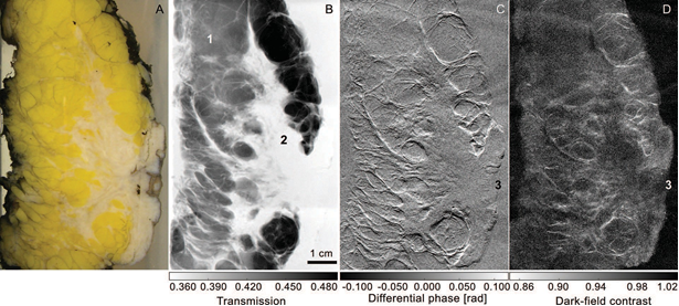
The photograph (A) shows the mastectomy specimen. Images (B), (C), and (D) show multimodal x-ray projection images (transmission, di?erential phase- contrast and dark-?eld contrast, respectively) of the mastectomy sample resulting from stitching eight single images together. The images were acquired on a CLS that was installed at Lynceans Palo Alto facility.
E. Eggl, S. Schleede, M. Bech, K. Achterhold, S. Grandl, A. Sztrokay, K. Hellerhoff, D. Mayr, R. Loewen, R. Ruth, M. F. Reiser, and F. Pfeiffer. X-ray Phase-Contrast Tomosynthesis of a Human Ex Vivo Breast Slice with an Inverse Compton X-ray Source, EPL, 116 (2016) 68003 [doi.org/10.1209/0295-5075/116/68003]
Coronary Angiography
The ability to significantly reduce the contrast media in iodine and gadolinium based X-ray coronary angiography through the use of quasi-monochromatic spectra is demonstrated at the MuCLS.
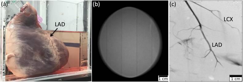
MuCLS angiography image. (a) Photograph of the sample in waterbath. (b) Empty image of full MuCLS beam. (c) Quasi-mono-energetic angiography image of a porcine heart acquired at the MuCLS, with iodine-based contrast agent injected into the left coronary artery. Visible are the left anterior descending artery (LAD) and the left circumflex artery (LCX).
E. Eggl, K. Mechlem, E. Braig, S. Kulpe, M. Dierolf, B. Günther, K. Achterhold, J. Herzen, B. Gleich, E. Rummeny, P. Noël, F. Pfeiffer, and D. Münzel. Mono-Energy Coronary Angiography with a Compact Synchrotron Source, Scientific Reports, 7:42211 (2017) [doi: 10.1038/srep42211]
Live Bio-Imaging
The potential for live bio-imaging research studies that capture biological function using short exposures is made possible by the partially spatially coherent, high flux X-ray beam generated at the MuCLS.
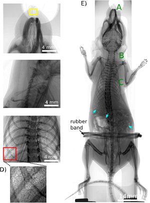 Panels (A)(C) show the interesting areas in lung and airway imaging in an ex-vivo mouse model. (A) The nasal airways.(B) Trachea region (for A and B detector pixel size = 6.5 ?m, exposure time = 1 s), (C) Lung (detector pixel size = 13 ?m, exposure time = 1 s), (D) is a magnification of the red box in (C) and shows the speckle pattern which results from the air sacs in the lungs, the alveoli. (E) Overview scan of the ex-vivo mouse imaged with propagation-based X-ray phase-contrast, composite image using 1 m propagation and 10 second exposure times (detector pixel size = 6.5 ?m)
R.Gradl, M. Dierolf, L. Hehn, B. Günther, A. Önder Yildirim, B. Gleich, K. Achterhold, F. Pfeiffer & K.S. Morgan. Propagation-based Phase-Contrast X-ray Imaging at a Compact Light Source, Scientific Reports, 7:4908 (2017) [doi:10.1038/s41598-017-04739-w]
Posted September 1, 2017