Correlative Microscopy
Correlative microscopy is commonly used to describe the combining of information from two or more different types of microscopy techniques. The concept allows the user to look at the same location on a sample using different microscopy techniques, with each technique yielding different information. By correlating the information, a more detailed understanding of a system can be derived that would otherwise not be possible.
Early Correlative Microscopy Techniques
Early correlative microscopy techniques involved time consuming manual relocating of samples under both types of microscope. This was at tedious process that risked time-based changes or contamination of sample to take place.
Advanced Correlative Microscopy Techniques
More recent advanced correlative microscopy techniques provide automated systems to ensure the same area of a sample is viewed by both microscopes. The ideal scenario has the sample located in situ so that it can be examined by several techniques simultaneously.
Raman Imaging and Scanning Electron Microscope RISE
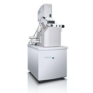
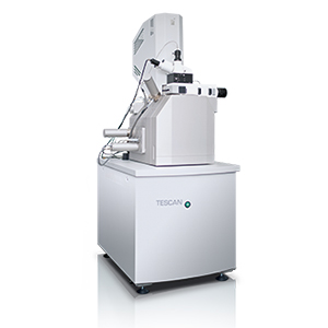
The RISE Microscope from TESCAN and WiTEC fully integrates a Scanning Electron Microscopy (SEM) and a Raman Microscope. This correlative microscopy technique combines the ultra-structural surface information provided by the SEM with the molecular compound information from the confocal Raman microscope.
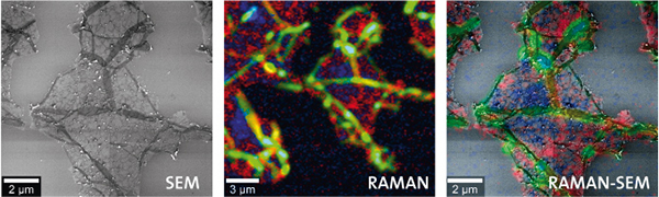
RISE Microscopy on graphene.
Left: SEM image of a graphene sample.
Middle: Color-coded confocal Raman image. The colors display the graphene layers and wrinkles. Image parameters: 20 ?m x 20 ?m, 150 x 150 pixels = 22,500 spectra, integration time: 0.05 s/spectrum.
Right: SEM image overlaid with the confocal Raman image.
Correlative Fluorescence and Scanning Electron Microscopy for Life Sciences
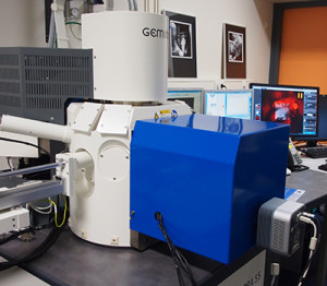
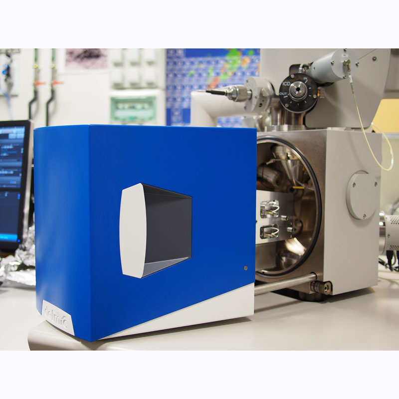
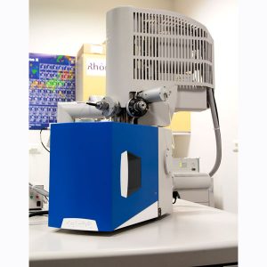
The innovative Delmic SECOM solution combines a fluorescence microscope built into the chamber of an SEM allowing the simultaneous imaging from both systems. This system can be retrofitted into existing SEMs and combines the functional information from the fluorescence microscope with the structural information from the SEM, making it ideal for the investigation of biological systems.
Correlative Cathodoluminescence and Scanning Electron Microscopy
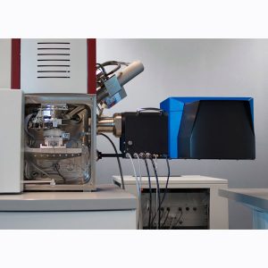
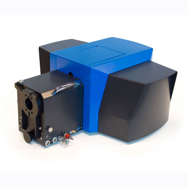
The Delmic SPARC system combines the analytical power of spectroscopic cathodoluminescence with SEM imaging. Spectroscopic cathodoluminescence provides valuable insights into materials types, purity and defects while at the same time offering optimal sensitivity, control and versatility. This is of specific interest to micro and nanophotonics.