
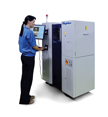
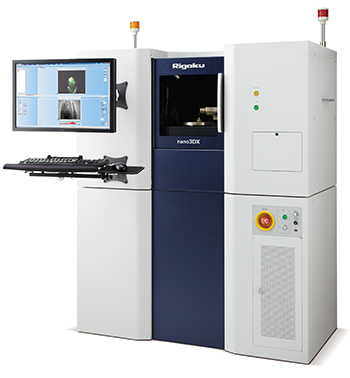
nano3DX – X-Ray Microscope
The nano3DX is a true x-ray microscope capable of high resolution microtomography of large samples. This is possible due to the rotating anode x-ray source and high resolution CCD imaging system.
Rotating Anode
The rotating anode gives the system the ability to change anode materials to produce x-rays at different wavelengths. This in turn allows the system optimise image quality via contrast and transmission to suit the materials being analysed.
Some examples of optimum radiation include:
- Chromium thin biological specimens and thin organic films
- Copper pharmaceuticals and carbon fibre reinforced plastics (CFRP)
- Molybdenum bone, silicates and aluminium composites
The rotating anode also provides fast data acquisition.
X-Ray Microscopy
In tomography, samples are studies as thin slices. Microtomography takes this a step further and each slice is thin enough so that it can be examined with an optical microscope. Classical tomography is both time consuming and tedious.
X-ray tomography visualises the entire sample at multiple rotation angles. The images are processed by a computer and sophisticated algorithms allow the individual slices to be reconstructed to form a 3D model that can in turn be sliced or viewed from any angle. This allows the user to probe the inner structure of an object in a non-destructive manner, providing valuable insights into its workings whether it be material, electronic or biological.
Resolution
The nano3DX operates with a voxel size of 0.27µm and a spatial resolution in the 0.6 to 0.8µM range. While some other instruments can match this resolution in a small scale, the nano3DX can offer this for larger samples with a much larger field of view.
Sample Size
The nano3DX is capable of measuring samples 14.4 x 10mm at a 4 ?m pixel size in a single scan, producing an image 3300×2500 pixels.
What Information Does an X-Ray Microscope Yield?
X-ray microscopy can indicate changes in composition and density. This can reveal such things are cracks or voids, in particular those beneath the surface. Furthermore, it can be used to determine such things are particle size distributions, fibre orientation as well as porosity and pore connectivity.
Key Features
- 3 x-ray wavelengths (Copper, chromium and molybdenum) for optimal imaging of different samples
- Ultra-wide field of view, 25X larger than similar systems
- Parallel beam geometry for high contrast and rapid date collection
- Auto 5-axis (XYZ and rotation) stage and on-axis imaging
- Produces high resolution 3D images
- High power (1.2kW) rotating anode x-ray source
- High contrast for low atomic number materials
- High resolution CDD imager
 Download the AXT NDT Product Guide
Videos produced using microtomography data generated by a Rigaku nano3DX x-ray microscope.