Microdiffraction – XRD Analysis of Small Areas
Microdiffraction is a very exciting relatively modern experimental technique made feasible by recent advances in hardware and software. It can be termed of as the XRD analysis of small samples or areas, including mapping.
Enabling Hardware
X-Ray Source and Optics
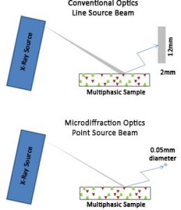 A critical piece of this experiment is to have a small monochromatic beam of intense X-rays and whilst this is readily available at the Synchrotron, it is now also possible to have small beams around 10µm in size in the homelab with modern day microfocus RAGs (Rotating Anode Generators), optics and collimation.
RAG’s have the advantage that they generate several times more flux compared to more conventional sealed X-ray tube systems. This means that there is much more X-ray intensity available for analysis enabling the detection of trace elements and faster analysis times, especially in conjunction with tailored optics that focus the X-rays down to tiny areas.
Detectors
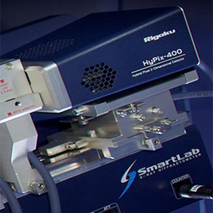
In addition, it is extremely advantageous to have a large and sensitive 2D detector that is capable of capturing the event in question in a single frame and then converting a 2D Debye-Scherrer representation by means of integration into a conventional plot of angle vs. intensity.
Initially photographic film was used as a detection method and this was in recent times superseded by imaging plate detectors. We have now reached an exciting point in detector technology with the introduction of Hybid Pixel Array Detectors (HPAD). Such detectors are 2D with an almost infinite dynamic range attributable to the extremely fast millisecond readout times, they are light, compact and robust in design. HPADs provide direct detection of X-rays are single photon counting with an unsurpassed signal to noise (zero dark noise and read noise).
Systems
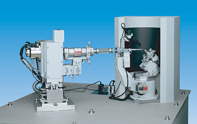
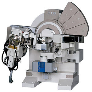
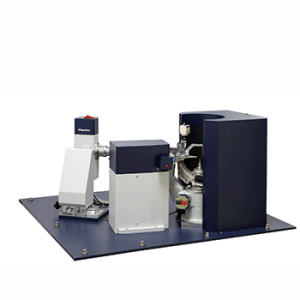
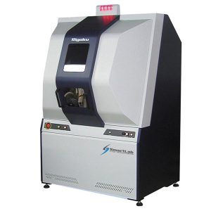
Rigaku offer two types of system that can perform microdiffraction:
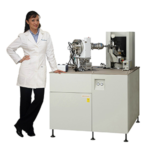
- Dedicated microdiffractometers e.g. the Rapid II with RAG, Imaging Plate Detector and industry-leading small focal spot size (down to 10µm)
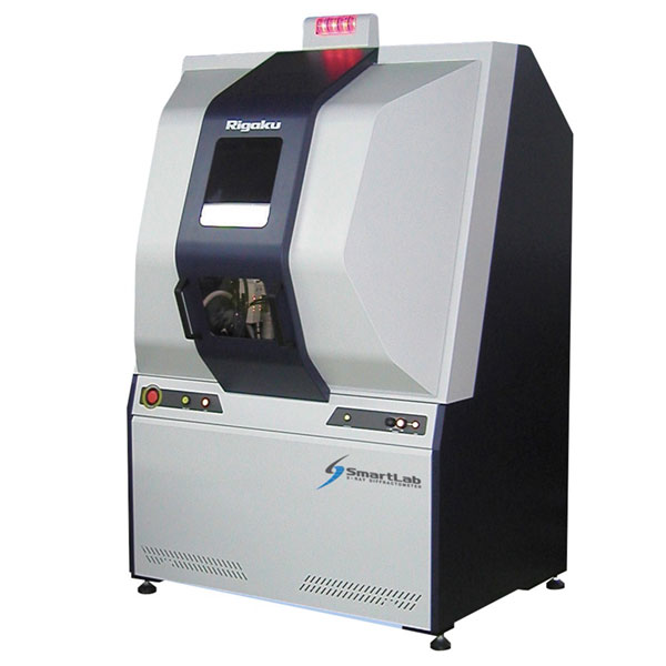
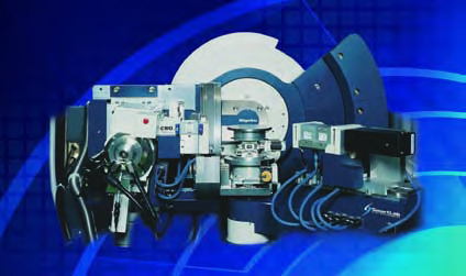
- Highly versatile diffractometers e.g. the 9kW SmartLab with RAG, also able to run XRD is a range of other configurations
Applications of Microdiffraction
Microdiffraction is particularly favoured where the region of interest is too small for conventional XRD techniques. It is also suited to analysis of valuable samples and can be used as a form of non-destructive testing and verification.
A single area may be of specific interest or a series of regions, and one can raster over larger regions by point mapping to collect numerous diffraction patterns. This is commonly known as data mapping (DM) and is of great interest to the geological/archaeological, materials and innovative manufacturing sectors.
In addition to conventional XRD information (phase identification for compound identification), the information revealed by way of the data, diffraction function map (DFM) probes into specific properties such as:
- Stress/strain
- Crystallinity
- Crystallite size
- Texture
Microdiffraction is commonly used in applications including:
- Geology e.g. inclusion identification
- Archaeology
- Test pads on patterned wafers
- Compound libraries formed by combinatorial chemistry
- Failure analysis
- Quality control in manufacturing
Related Products