FIB-SEM platform with in-situ ToF-SIMS for Geological Sample Preparation and Advanced Analysis
By Will Rickard1 and Cameron Chai2
1. John de Laeter Centre, Curtin University
2. AXT Pty Ltd
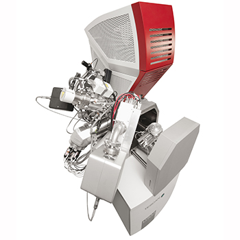
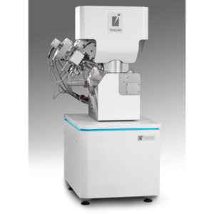
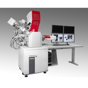
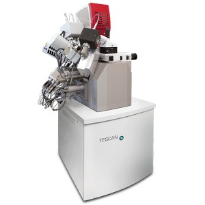
In 2015, Curtin University installed a TESCAN LYRA Ga-FIB-SEM with a fully integrated Time of Flight Secondary Ion Mass Spectometer (ToF-SIMS). This system was purchased primarily for sample preparation for their atom probe which is dedicated to geoscience research. At the time, this was the first installation of its type in Australia.
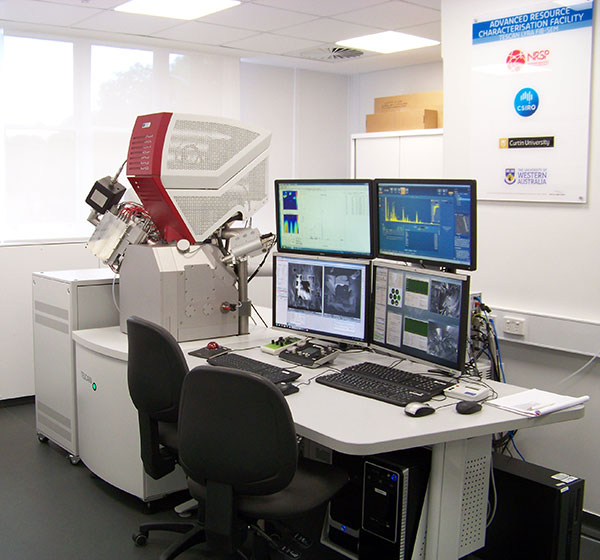
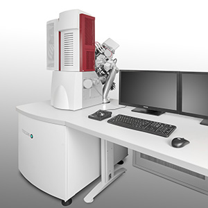
Figure 1. TESCAN LYRA FIB-SEM with integrated ToF-SIMS at Curtin University.
While many researchers use electron imaging in conjunction with microanalytical techniques such as EDS or EBSD to identify areas of interest to prepare atom probe samples, Curtin Universitys FIB-SEM facility leader Dr. Will Rickard has additionally used in situ ToF-SIMS. TOF-SIMS offers a number of advantages for targeting fine variations in chemistry when compared to X-ray based analytical techniques such as EDS. These include:
- Improved spatial resolution (lateral resolution <50nm, depth resolution <20nm)
- Improved sensitivity (down to ppm levels for some elements)
- Improved light element detection (Li is routinely detectable)
- Isotopic information
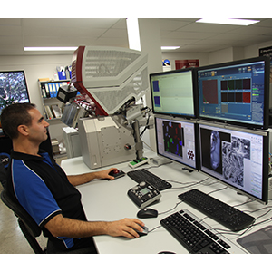
The FIB-ToF-SIMS technique has enhanced our microanalytical capability and improved our work flow for atom probe sample preparation. ToF-SIMS elemental mapping can offer spatial resolution an order of magnitude better than techniques like EDS, which allows us to more accurately pinpoint areas of interest for atom probe analysis such as low angle boundaries or other areas where there are fine changes in the chemistry. This is important as the atom probe typically measures sample volumes of the order of 0.01µm3 and using the ToF-SIMS enables us to accurately target fine structures, thus better utilising time on the atom probe. Having the TOF-SIMS and FIB co-located in the same vacuum chamber also allows us to prepare samples more quickly, explained Dr. Rickard.
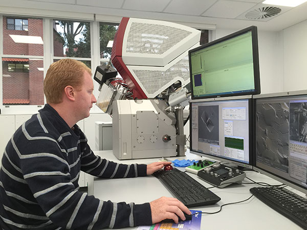
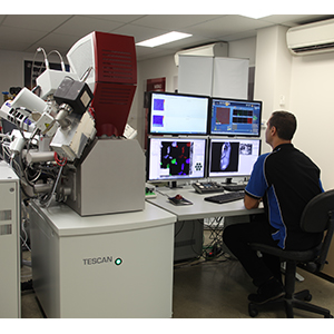
Figure 2. Dr. Will Rickard operating the TESCAN LYRA FIB-SEM.
The FIB and ToF-SIMS combination also allows researchers at Curtin University to build 3 dimensional elemental maps. Using the FIB, they can simultaneously remove a layer of material and analyse the material that has been removed using the ToF-SIMS, obtaining datasets in just a few minutes. The TESCAN FIB-ToF-SIMS system that Curtin have is also equipped with EDS and EBSD. By correlating datasets from these techniques with the atom probe results, the researchers can have defined a robust and reliable analytical workflow that provides a detailed characterisation across a range of length scales.
To date, the main areas where the FIB-ToF-SIMS and atom probe have made the biggest impact are:
- Analysis of zircon and other accessory phases where it has been used to study the trace element chemistry over a wide range of length scales.
- Analysis of Li bearing minerals in combination with automated mineralogy and laser ablation.
- Solid Oxide Fuel Cells (SOFC) research where it has been used for high resolution elemental mapping and depth profiling.
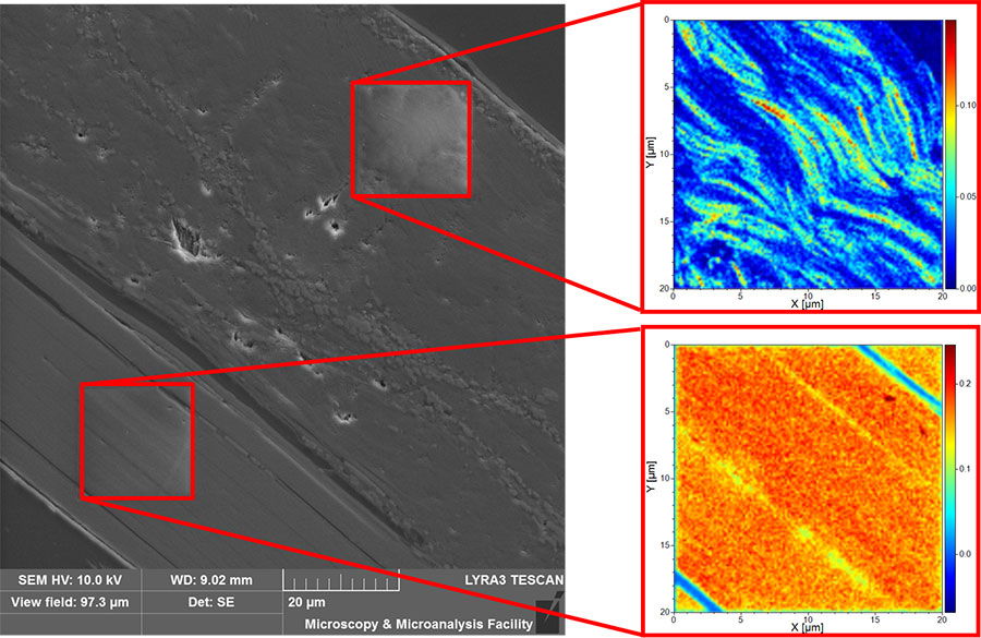
Figure 3. Poassium distribution in biotite.
One area where the system has been a revelation is the analysis of lithium-bearing minerals. Lithium is very difficult to detect using x-ray techniques such as EDS due to its low energy characteristic x-ray emission. While it can be detected by laser ablation and bulk chemical analyses, these techniques are incapable or limited in providing spatially resolved data. The FIB-ToF-SIMS overcomes these issues and as such as been attracting the attention of industrial partners who are looking to take advantage of the technology benefits. Dr. Rickard will be presenting this work at the upcoming ALTA Metallurgical conference that will be held from May 20th to 27th in Perth.
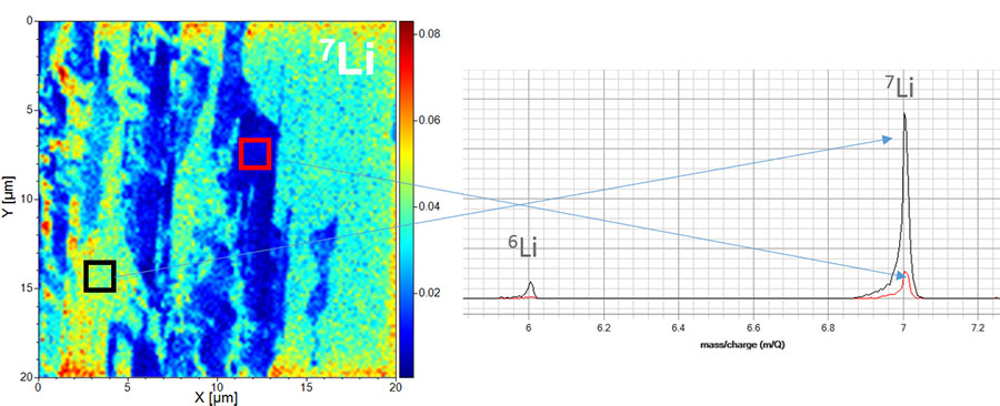
Figure 4. Figure 4. Lithium variation in a mica sample. Lithium is not detectable using conventional EDS.
For more information about the TESCAN LYRA and the ARCF atom probe visit www.geoscienceatomprobe.org
Note: This article was previously published in the AMMS Newsletter.