
Multi-Modal X-Ray Microscope and Micro-CT with Bragg Magnifier Based on Rigaku X-Ray Generator
Dr. Daniele Pelliccia from RMIT University has built Australias most versatile X-ray instrument. In conjunction with Patrik Vagovic and other researchers from Germany and the Slovak Academy of Sciences , Daniele has recently published a paper entitled Laboratory-Based Multi-Modal X-ray Microscopy and Micro-CT with Bragg Magnifier in the journal Optics Express (DOI: 10.1364/OE.23.018391). Using novel Bragg magnifier optics, the researchers have been able to combine different microscopy modalities together into a single instrument using a lab-sized X-ray source as opposed to resorting to synchrotron radiation.
At the heart of the system is a Rigaku FRE+ rotating anode X-ray generator high brightness X-ray source. Out of the box, the FRE+ has a focal spot size in the order of 80 to 100µm and is not generally suited used imaging applications. However, Daniele and his colleagues designed a novel optics system that magnifies the X-ray beam acting as X-ray zoom lens, resulting in an increase in resolution. While the optic is somewhat complex, it actually adds an additional level of flexibility to the system.
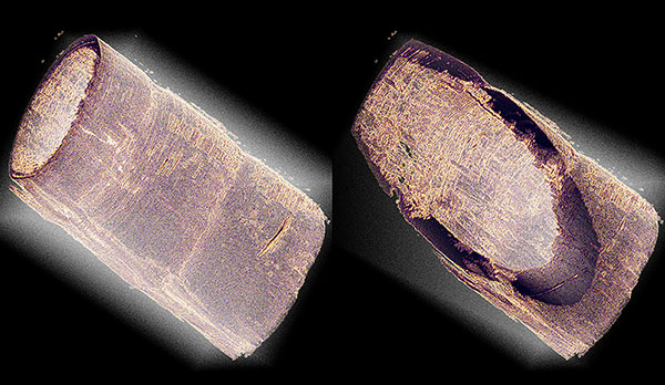
Selected 3D renderings of the micro-CT of the antenna of a tiger prawn. The specimen is about 1 mm in diameter and produces a significant attenuation for X-rays. Micro-CT enables non-destructive inspection of sample volumes. In this example both outer and inner walls as well as he structure of the antenna wall itself can be studied through virtual cuts across the rendered volume.