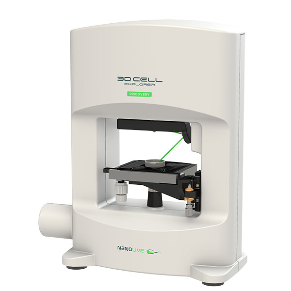
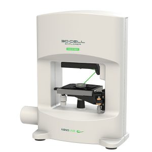
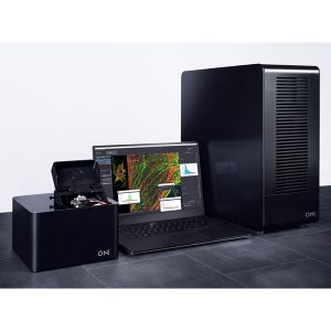
3D Cell Explorer Discovery Holo-Tomographic Microscope for Teaching
The 3DCX-Discovery is based on the award-winning Nanolive 3D Cell Explorer. It has been designed for teaching environments, is priced attractively for schools and universities and is capable of delivering high-quality images with nanometric resolution in just seconds.
The 3DCX-Discovery is a plug-and-play system that requires little if any sample preparation. Furthermore, its operation is simplified by the fact that it does not need any markers, stains or dyes. As such it is the ideal instrument for introducing students to live cell imaging and cell biology.
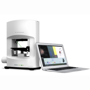
This particular model is aimed at secondary and tertiary teachers and lecturers looking to take their teaching to the next level. The affordability of the 3DCX-Discovery enables students to physically and dynamically observe and analyse cells rather than just looking at old, boring, static images on a screen. It is perhaps the ultimate teaching tool to stimulate biology classes – to sit up, take notice and get involved.
What You Can Do with It
 The 3DCX-Discovery is a great tool allowing you to:
- Observe cells with incredible resolution
- Observe a vast array of different cells
- Characterise different cellular structures
- Explore single cells and cell cultures stain-free
- Instantly image cells
- Observe cell-cell interaction
- Observe cellular responses to different stimuli
- Observe cells without interfering with the natural physiology
Benefits of the 3DCX-Discovery
Some of the benefits of the 3DCX-Discovery that make it ideal for teaching applications include:
- Analysis of the true cells with no labels
- Ability to observe cells in their natural environment
- Realtime imaging
- The ability to measure cell dimensions and volumes
How Does It Work?
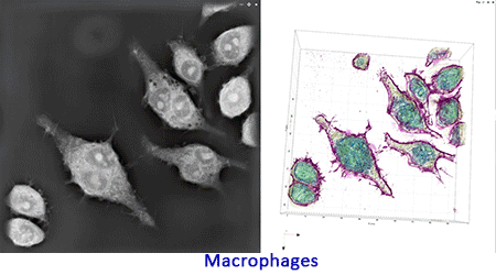
The 3DCX-Discovery shines a laser through your specimen. Using holographic tomography, it takes 96 slices through a 30µm thick sample and differentiates different cellular structures such as nuclei, vesicles etc. based on changes in refractive index with a resolution of better than 200nm
The slices are then reconstructed to create a 3D image of your cell, much like an MRI system.
The entire measurement takes less than a second, enabling you to image in real time and observe changes as they happen. You can also view each individual slice in great detail.
Digital Staining
Rather than using chemical stains or dyes (and the time-consuming associated sample preparation) to highlight different structures, the 3DCX-Discovery allows you to use an offline version of the STEVE software to digitally stain your cells.
This has the benefit of freeing the instrument up for taking measurements, and allowing staining to be carried out at a later time.
Get Involved
To get involved, simply contact AXT!