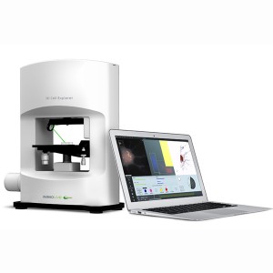
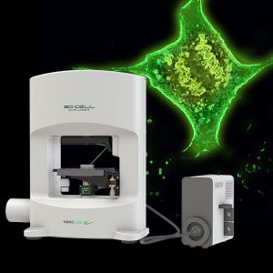
GLIM Gradient Light Interference Microscopy for Life Science – QPI
Quantitative Phase Imaging of specimens over 350µm thick
Gradient Light Interference Microscopy or GLIM is a new quantitative phase imaging (QPI) technique. The technology exploits a special case of low-coherence interferometry to extract phase information from 2D and 3D specimens. This can be used to determine:
- Cell mass
- Cell volume
- Surface area
- Evolution of cells over time
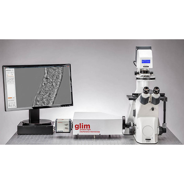
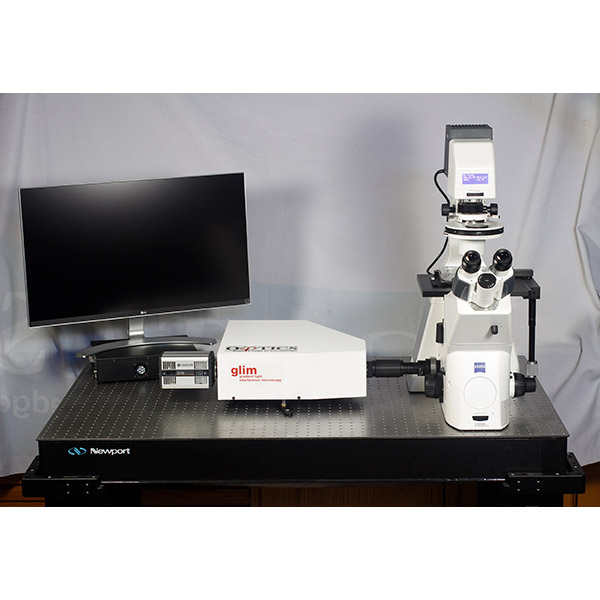
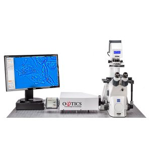
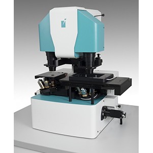
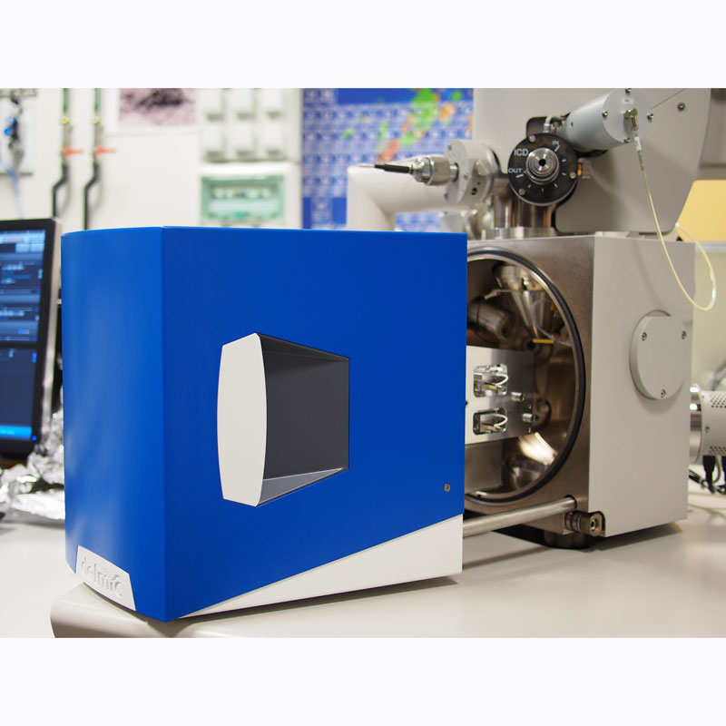
GLIM is supplied as a module and is a simple upgrade to your existing inverted DIC (differential Interference Contrast) microscope It provides it with additional analytical capabilities, as well improving its contrast, thus enabling QPI.
Key Capabilities
- Non-invasive: no sample preparation. (Resolves samples with thickness from 50 µm 350 µm+)
- Quantitative measurements: thickness and dry mass
- Label free continuous imaging from milliseconds to days
- Integrates with existing research grade microscopes
- Programmed 4D (tiling, z-scan, time series) scanning and acquisition at up to 12fps with full camera resolution
- Multichannel imaging (including fluorescence channels) with seamless overlay
- ImageJ-based toolkit for measurement and 3D image rendering
What is GLIM
GLIM is a non-invasive phase imaging technology that works by quantifying the optical path difference in biospecimens and converts them into thickness, dry mass, density and refractive index maps.
How Does GLIM Work?
The contrast in optical imaging of thick specimens is controlled by multiple scattering limits. GLIM works by suppressing the incoherent background brought about by multiple scattering. It achieves this by combining multiple intensity images that correspond to controlled phase shifts between two interfering waves
When a live tissue culture is imaged using DIC, two interfering fields are generated. They are identical except for a small transverse spatial shift. This geometry ensures that the two fields suffer equal degradation as a result of multiple scattering. Through accurate control; of the phase shift between the two waves, GLIM generates multiple intensity images with the same incoherent background, but different coherent contributions. This enables GLIM to filter out much of the contributions form multiple scattering. This produces high contrast images of thick objects.
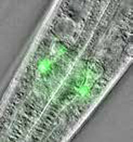 | 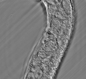 | 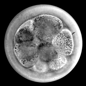 |
| Phi Optics GLIM + GFP overlay image of pharynx | GLIM image of live C.Elegans | GLIM image bovine embryo |
GLIM can also produce tomographic images of thin and thick specimens using optical sectioning when the illumination condenser if fully open.
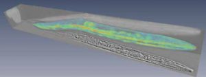 | 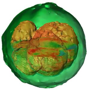 |
| 3D reconstruction of C.Elegans. | Bovine embryo 3D reconstruction using GLIM data. |
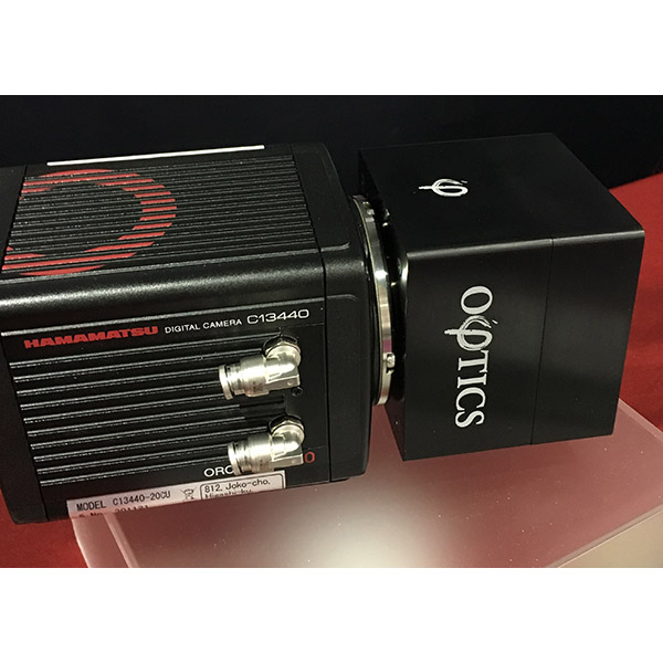
How Does GLIM Integrate with your Light Microscope?
The Phi Optics GLIM module simply attaches to the C-mount of your existing inverted DIC microscope. The module includes the CCD camera.
Applications
- Brain section
- 3D organoids
- Multi-cellular organisms e.g. C. Elegans, C. Planaria
- Embryos
- Animal models e.g. worms, zebrafish, fruit flies